
Health
How to Decide If You Need Penile Enlargement or a P-Shot in Bournemouth Based on Your Goals

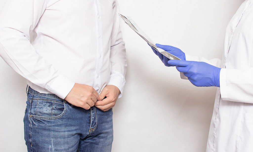
Men researching intimate health treatments may have different concerns, from wanting to change penile size to seeking improvements in erectile function or sexual confidence. Understanding these goals is an important first step when comparing penile enlargement in Bournemouth with the Priapus Shot, commonly called the P-Shot.
Although both treatments are associated with men’s intimate health, they have different purposes and should not be viewed as interchangeable procedures. Penile enlargement focuses on changing penile appearance or dimensions, depending on the technique used. The P-Shot is a platelet-rich plasma (PRP) treatment intended to support tissue health, although evidence for its effectiveness in treating erectile dysfunction or increasing penile size remains limited.
Choosing between these options requires an understanding of your expectations, medical history, potential risks and the available clinical evidence. A consultation with a qualified medical professional can help determine whether either treatment is appropriate for your circumstances.
Understanding the Difference Between Penile Enlargement and the P-Shot
Before making a decision, it is helpful to understand what each treatment involves and what it is intended to address.
What Is Penile Enlargement?
Penile enlargement refers to procedures intended to change penile dimensions or appearance. Depending on the clinic and treatment method, this may involve injectable dermal fillers or surgical approaches.
Non-surgical enlargement using hyaluronic acid-based fillers may increase penile girth by adding volume beneath the skin. The amount of change, duration of results and suitability depend on the product, technique and individual anatomy.
Surgical procedures may involve different approaches and carry different recovery requirements and risks. These should be discussed separately with an appropriately qualified surgeon.
Men should understand that enlargement treatments do not necessarily improve erectile function, sexual stamina or sexual satisfaction. Their primary purpose is to address physical appearance or dimensions.
What Is the P-Shot?
The P-Shot uses platelet-rich plasma taken from the patient’s own blood. A sample is collected and processed to concentrate platelets before the preparation is injected into selected areas of the penis.
Platelets contain growth factors involved in the body’s natural healing processes. The treatment is promoted by some providers as an option for supporting penile tissue health and sexual function.
However, clinical evidence supporting the P-Shot for erectile dysfunction, penile enlargement or other sexual health concerns is currently limited. It should not be presented as a proven treatment or a guaranteed alternative to established medical therapies.
Identify Your Main Treatment Goal
The most useful way to compare these treatments is to identify the concern you want to address. Your primary goal can influence which options a clinician recommends.
If Your Priority Is Increasing Penile Girth
Men who want to change penile girth may research enlargement procedures, including injectable treatments. These approaches are designed to alter physical dimensions rather than directly treat erectile dysfunction.
A medical assessment should establish whether the requested change is realistic and whether the proposed technique is appropriate. It should also cover the possibility of uneven results, lumps, swelling, infection and other complications.
It is important to avoid making decisions based solely on photographs or advertised measurements. Individual results vary, and no procedure can guarantee a particular outcome.
If Your Concern Is Erectile Function
If your main concern is difficulty achieving or maintaining an erection, the first step should be a medical assessment rather than an appearance-focused procedure.
Erectile dysfunction can have several causes, including cardiovascular conditions, diabetes, hormonal changes, medication effects, stress and psychological factors. Identifying the underlying cause is important because treatment should address the relevant health issue.
The P-Shot is sometimes promoted for erectile function, but there is not enough high-quality evidence to establish it as a reliable treatment. Established options, including lifestyle changes, prescribed medication and other medical interventions, may be more appropriate depending on the diagnosis.
If You Want to Address Confidence or Body Image Concerns
Concerns about penile appearance can affect confidence and relationships. However, the emotional impact of these concerns should be considered alongside the physical treatment options.
A clinician can discuss normal anatomical variation, realistic expectations and whether a procedure is likely to address the concern. If anxiety about size or appearance is persistent or significantly affects daily life, psychological support may also be beneficial.
A treatment should not be chosen simply because it is advertised as a way to improve confidence. Its likely benefits and limitations need to be understood first.
Comparing the P-Shot in Bournemouth With Enlargement Treatments
Men researching a P-shot in Bournemouth should understand that PRP treatment and penile enlargement address different concerns. Comparing them based on the intended outcome can help prevent unrealistic expectations.
Treatment Purpose
Penile enlargement procedures aim to change physical dimensions or appearance. The P-Shot is intended to use platelet-rich plasma to support tissue processes, although its clinical benefits for sexual function remain uncertain.
Neither treatment should automatically be considered a solution for all sexual health concerns.
Procedure and Preparation
Penile enlargement procedures vary according to the technique used. Injectable treatments generally involve an assessment, preparation of the treatment area and carefully administered injections. Surgical approaches involve a different level of intervention and may require more extensive preparation and recovery.
The P-Shot involves drawing a blood sample, processing it to obtain platelet-rich plasma and administering the preparation through injections.
Both procedures require appropriate medical assessment, sterile techniques and clear explanations of potential complications.
Recovery and Aftercare
Recovery depends on the treatment method, individual health and the clinician’s instructions.
After injectable enlargement, patients may experience temporary swelling, bruising, tenderness or changes in the treated area. Surgical procedures may involve additional restrictions and a longer recovery period.
Following a P-Shot, temporary discomfort, swelling or bruising may occur around the injection sites. Patients should follow the provider’s aftercare instructions and seek medical advice if they experience severe pain, significant swelling, fever or other concerning symptoms.
Important Factors to Consider Before Choosing a Procedure
A treatment decision should involve more than comparing the advertised benefits. Several medical and practical factors deserve attention.
Your Medical History
Existing medical conditions, allergies, previous procedures and current medication can influence treatment suitability.
For example, bleeding disorders, active infections and certain medications may affect whether an injectable procedure is appropriate. Patients should provide complete and accurate medical information during their consultation.
The Evidence Supporting the Treatment
Ask the clinician what scientific evidence supports the proposed procedure and whether the expected benefits have been demonstrated in well-designed clinical studies.
This is particularly important when discussing the P-Shot, as promotional claims may be stronger than the available evidence.
Potential Risks and Complications
Every medical procedure carries potential risks. Injectable treatments may cause infection, bruising, swelling, discomfort, uneven results or tissue complications. More serious complications can occur, depending on the procedure and technique.
Ask what complications are possible, how frequently they occur, and what arrangements are available if treatment-related problems develop.
Costs and Long-Term Expectations
Treatment costs may depend on the technique, amount of product used, clinician’s fees and any follow-up appointments.
Patients should ask whether additional sessions or maintenance treatments may be required. They should also understand that results may change over time and that further procedures are not always advisable.
Questions to Ask During Your Bournemouth Consultation
A consultation provides an opportunity to understand the proposed treatment and decide whether it is suitable.
Consider asking the following questions:
- What is the main purpose of the recommended treatment?
- Am I medically suitable for the procedure?
- What results are realistic in my circumstances?
- What clinical evidence supports the treatment?
- What are the possible short-term and long-term complications?
- Who will perform the procedure, and what qualifications do they hold?
- What aftercare and follow-up appointments are included?
- What should I do if I experience an unexpected complication?
A reputable provider should answer these questions clearly and allow enough time for you to make an informed decision. You should not feel pressured to book a procedure during your initial appointment.
When Neither Treatment May Be the Right Option
Not every concern requires an enlargement procedure or PRP injection. If you are experiencing erectile difficulties, pain, changes in penile shape or other symptoms, a medical assessment should come first.
Conditions such as Peyronie’s disease, hormonal disorders and cardiovascular problems may require specific investigations and established treatments. An appearance-focused procedure may not address the underlying issue.
Similarly, if your expectations involve a substantial or guaranteed increase in size, it is important to discuss the limitations of available procedures before proceeding.
In some circumstances, monitoring, lifestyle changes, medication or specialist referral may be more appropriate than an injectable treatment.
Conclusion
Deciding between penile enlargement and the P-Shot in Bournemouth begins with understanding your personal goals and the differences between the procedures. Enlargement treatments are intended to change penile appearance or dimensions, while the P-Shot uses platelet-rich plasma and has limited evidence supporting its promoted benefits for sexual function.
Before proceeding, consider your medical history, treatment expectations, available clinical evidence, potential complications and recovery requirements. A consultation with a suitably qualified medical professional can help you understand the options and identify an appropriate course of action.
Ultimately, the best decision is not necessarily the procedure with the most appealing claims. It is the option that is medically appropriate, supported by realistic expectations and aligned with your individual health needs.
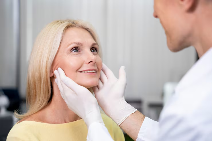
Recovery after combined facial procedures requires careful planning, patience, and close adherence to your surgeon’s instructions. When a deep plane neck lift in Manchester is performed alongside a midface procedure, the recovery plan needs to account for healing in more than one area. Swelling, bruising, tightness and temporary changes in sensation can occur, and the timing of recovery varies between patients.
The aim of this guide is to explain what to expect, how to prepare your home and which habits can support a safer and more comfortable recovery. Your surgeon’s personalised advice should always take priority over general guidance.
1. Understand the Recovery Process Before Surgery
Combining procedures can mean recovering from changes to different facial areas at the same time. A deep plane neck lift addresses deeper tissues in the neck and jawline, while a midface lift repositions supporting tissues around the cheeks and lower eyelid area. Each area has its own healing considerations.
The neck may feel tight, swollen or temporarily numb. The cheeks and lower eyelids may also develop bruising and swelling, with a firm sensation as repositioned tissues settle. These effects are generally part of the early healing process, but your surgical team should explain which symptoms are expected in your circumstances.
Discuss Your Individual Recovery Plan
Before surgery, ask your surgeon how the combined procedures may affect your recovery, when dressings or any drains should be reviewed, and when you can resume everyday activities. Arrange follow-up appointments and make sure you have a contact number for questions or concerns after discharge.
2. Prepare Your Home and Arrange Practical Support
Preparing your home before surgery can make the first few days more manageable. You may feel tired, have limited mobility and need help with ordinary tasks while the effects of anaesthesia and surgery wear off.
Create a Comfortable Resting Area
Choose a quiet place where you can rest without repeatedly climbing stairs or reaching for essential items. Keep your telephone, prescribed medication, water, tissues and surgeon-approved supplies nearby.
Your surgical team may recommend sleeping with your head elevated to help manage swelling. Arrange suitable pillows or a supportive resting arrangement in advance, and follow the specific positioning instructions you receive.
Arrange Help With Daily Activities
Ask a trusted adult to accompany you home and stay with you for the period recommended by your surgical team. You may need assistance with meals, shopping, household tasks and transport to follow-up appointments.
Do not plan to drive yourself home after surgery. Your ability to drive again will depend on your recovery, medication and surgeon’s approval.
3. Follow Your Wound Care and Medication Instructions
Good aftercare helps protect healing tissues and reduces the risk of avoidable complications. Your surgeon will explain how to care for incision sites, whether dressings need changing and when you can wash your hair or face.
Keep Incisions Clean and Protected
Follow the instructions provided for cleaning and protecting surgical wounds. Avoid rubbing, pulling or applying unapproved creams and skincare products near the incision sites. Do not remove dressings, stitches or surgical supports unless your team tells you to do so.
If you notice increasing redness, unusual discharge, worsening discomfort or a wound that appears to be opening, contact your surgical team promptly.
Take Medication as Prescribed
Use prescribed pain relief and other medication according to the instructions given. Do not add over-the-counter medicines, supplements or herbal products without checking with your surgeon or pharmacist, as some can affect bleeding or interact with prescribed treatments.
4. Manage Swelling, Bruising and Discomfort
Swelling and bruising are common after facial surgery. When more than one area has been treated, these effects may be noticeable around the neck, jawline, cheeks and lower eyelids.
Your surgeon may recommend specific measures to manage discomfort and swelling. Follow their advice about cold compresses, head elevation and activity. Do not apply ice directly to the skin or place pressure on operated areas unless specifically instructed.
Be Patient With Changes in Appearance
Swelling can make the face appear uneven during the early stages of healing. Temporary asymmetry does not necessarily indicate a poor result, as each side may settle at a different rate.
Avoid judging the outcome too soon. The tissues need time to heal, soften and adapt to their repositioned position. Your surgeon can explain the expected recovery milestones for your particular procedures.
5. Protect Your Midface and Neck During Everyday Activities
A combined recovery requires care when moving, sleeping and returning to normal routines. Avoid activities that stretch the neck, place pressure on the cheeks or increase blood pressure during the initial healing period.
Keep Your Movements Gentle
Follow your surgeon’s advice about walking and light movement. Short, gentle walks may be encouraged when appropriate, but strenuous exercise, heavy lifting and activities that involve bending or sudden movements should be avoided until you are cleared.
Do not massage the cheeks, neck or jawline unless your surgeon specifically recommends it.
Follow Advice About Sleeping and Positioning
Your surgeon may advise sleeping on your back with your head elevated. Avoid sleeping face down or placing pressure on the operated areas. Ask when it is safe to return to your usual sleeping position.
6. Support Healing Through Nutrition and Healthy Habits
Your body needs adequate energy and nutrients to repair tissue after surgery. Aim for balanced meals containing protein, vegetables, fruit and other nutritious foods. If chewing feels uncomfortable, ask your team whether softer foods are suitable during the first few days.
Stay hydrated unless you have been given different medical instructions. Avoid smoking and vaping, as nicotine can impair circulation and interfere with wound healing. Follow your surgeon’s advice about alcohol and any restrictions involving medication.
7. Know When to Seek Medical Advice
Although swelling, bruising and tightness can be expected, certain symptoms require prompt assessment. Contact your surgical team if you experience increasing pain, worsening swelling, fever, spreading redness, unusual wound discharge or sudden changes in sensation or movement.
Seek emergency medical attention for severe breathing difficulty, chest pain, sudden significant bleeding or other symptoms that feel immediately dangerous. Do not wait for a routine follow-up appointment if you are concerned about a serious complication.
8. Attend Follow-Up Appointments and Return to Activities Gradually
Follow-up appointments allow your surgeon to assess incision healing, swelling and the position of the treated tissues. They also provide an opportunity to discuss concerns and receive advice about the next stage of recovery.
For patients undergoing midface lift surgery alongside a neck lift, the recovery schedule should account for both procedures. The clinic’s published guidance describes swelling and bruising during the first one to two weeks after midface lifting, while recovery following a deep plane neck lift can take longer. These are general estimates, not guarantees for combined surgery.
Return to Work and Exercise Only When Cleared
Some people may be ready for desk-based activities before they feel comfortable attending social events or undertaking physical work. Your surgeon will advise when you can return to work, drive and exercise. Avoid rushing back to strenuous activities simply because the initial swelling has improved
Conclusion
Recovering from a deep plane neck lift and midface lift performed together involves looking after multiple healing areas, following wound care instructions and allowing enough time for swelling to settle. Preparing your home, arranging practical support, taking medication as prescribed and attending follow-up appointments can make the process more manageable.
Most importantly, remember that recovery is individual. Follow your surgeon’s advice, report unexpected symptoms promptly and allow your tissues time to heal before judging the outcome. A careful, patient approach is an important part of supporting a safe recovery.
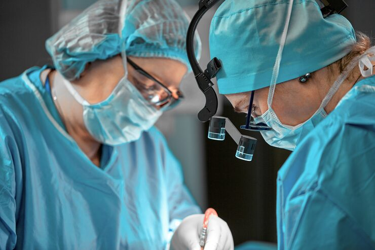
Salivary gland problems can cause discomfort, swelling, and recurring pain, particularly when saliva cannot flow normally through the ducts. For patients experiencing these symptoms, consulting London head and neck surgery specialists can help identify the underlying cause and determine an appropriate treatment approach. One diagnostic technique used in selected cases is sialendoscopy, a minimally invasive procedure that allows specialists to examine the inside of the salivary gland ducts.
Sialendoscopy provides a direct view of the duct system, helping clinicians investigate conditions such as salivary stones, duct narrowing, and inflammation. Unlike conventional imaging alone, this technique allows specialists to inspect the affected area using a small endoscope.
Understanding how the procedure works, when it may be recommended, and what patients can expect can help individuals prepare for a consultation with a qualified specialist.
What Is Sialendoscopy?
Sialendoscopy is a minimally invasive technique used to examine the ducts that carry saliva from the salivary glands into the mouth. It involves inserting a very thin endoscope equipped with a light and camera into a natural opening of the salivary duct.
The instrument allows the specialist to view the duct’s internal structure and identify abnormalities that may be responsible for a patient’s symptoms.
Understanding the Salivary Glands
The body has three major pairs of salivary glands:
- Parotid glands: Located in front of and below the ears.
- Submandibular glands: Found beneath the lower jaw.
- Sublingual glands: Situated beneath the tongue.
These glands produce saliva, which helps lubricate the mouth, support swallowing, and begin the digestive process. Saliva travels through small ducts before entering the oral cavity.
When a duct becomes blocked or narrowed, saliva may accumulate inside the gland. This can lead to swelling, discomfort, and sometimes infection.
How the Endoscope Works
The endoscope used during sialendoscopy is designed to pass through the natural salivary duct opening. Its small camera transmits images to a monitor, allowing the specialist to examine the duct’s interior.
Depending on the clinical findings, the procedure may also allow the specialist to investigate an obstruction more closely or perform certain treatments during the same session.
Why Specialists Use Sialendoscopy for Diagnosis
Salivary gland symptoms can have several possible causes. Swelling around the jaw, discomfort during meals, and repeated episodes of inflammation may indicate an issue involving the salivary ducts.
Sialendoscopy helps specialists investigate these symptoms by providing a direct view of the duct system.
Identifying Salivary Stones
Salivary stones, also known as salivary calculi, are hardened deposits that develop within the salivary glands or their ducts. They can obstruct saliva flow and cause swelling or pain, particularly when eating.
During sialendoscopy, a specialist may be able to identify the location and appearance of a stone. This information can help determine whether it may be suitable for removal using an endoscopic technique or whether another approach is necessary.
Detecting Duct Narrowing
Salivary ducts can become narrowed due to inflammation, scarring, or other structural changes. This condition, known as duct stenosis, may restrict saliva movement and contribute to recurring symptoms.
Sialendoscopy allows the specialist to examine the affected section and assess the extent of the narrowing. The findings can help guide decisions about further investigation or treatment.
Investigating Recurrent Inflammation
Repeated salivary gland inflammation may be associated with blockages, duct abnormalities, or other underlying conditions.
Direct examination of the duct can provide additional information when symptoms continue despite initial management. However, sialendoscopy is not suitable for every patient, and the specialist must assess whether it is appropriate.
When Is Sialendoscopy Recommended?
Sialendoscopy may be recommended when a patient’s symptoms suggest an obstruction or abnormality within a salivary duct. It is particularly useful when further investigation is needed after a clinical examination or imaging assessment.
Patients experiencing recurring swelling, discomfort during meals, or repeated episodes of salivary gland inflammation may benefit from specialist evaluation.
Symptoms That May Require Investigation
Common symptoms associated with salivary duct problems include:
- Swelling around the cheeks, jaw, or beneath the tongue.
- Pain that becomes more noticeable during meals.
- Recurrent salivary gland infections.
- A feeling of pressure around the affected gland.
- Reduced saliva flow or persistent dry mouth.
These symptoms do not automatically indicate a condition that requires sialendoscopy. Other medical problems can produce similar signs, so an appropriate assessment is important.
The Role of Medical Imaging
Before recommending an endoscopic examination, specialists may request imaging tests such as ultrasound, computed tomography (CT), or magnetic resonance imaging (MRI).
These investigations can help identify stones, gland abnormalities, and other structural changes. The choice of imaging depends on the patient’s symptoms and clinical history.
Sialendoscopy may provide additional information when imaging does not fully explain the problem or when direct examination of the duct is required.
How Sialendoscopy in London Supports Specialist Assessment
Access to specialist evaluation is important when salivary gland symptoms are persistent or recurring. Patients researching sialendoscopy in London can learn more about the procedure and discuss whether it may be suitable for their particular condition.
A specialist assessment usually begins with a discussion of symptoms, medical history, and previous investigations. The clinician may examine the affected area and review any available imaging before recommending further tests or treatment.
Preparing for the Procedure
Preparation depends on the planned procedure and the type of anaesthesia required. Patients may receive instructions about eating, drinking, medication, and arrangements for travelling home.
It is important to disclose existing medical conditions, allergies, and prescribed medication during the consultation. Patients should also ask the specialist about expected recovery time and any restrictions following the procedure.
What Happens During Sialendoscopy?
The procedure may be performed under local or general anaesthesia, depending on the clinical circumstances.
Once the patient is appropriately prepared, the specialist gently introduces the endoscope through the salivary duct opening. The camera provides images of the duct, allowing the clinician to inspect its internal lining and identify abnormalities.
If a stone or narrowing is found, the specialist may discuss whether treatment can be performed during the same procedure. The exact approach depends on the location, size, and nature of the problem.
Benefits and Limitations of Sialendoscopy
Sialendoscopy offers several potential advantages for selected patients, although it is not appropriate for every salivary gland condition.
Direct Visual Examination
One of its main advantages is the ability to examine the inside of the salivary duct directly. This can help specialists identify abnormalities that may not be fully characterised through other investigations.
Minimally Invasive Approach
Because the endoscope is introduced through the natural duct opening, the procedure may avoid the need for larger external incisions in suitable cases.
It can also allow certain obstructions to be treated while preserving the affected salivary gland. However, treatment outcomes depend on the individual condition and the findings during the procedure.
Possible Risks and Complications
Although generally considered minimally invasive, sialendoscopy carries potential risks. These may include temporary swelling, discomfort, bleeding, infection, or injury to the duct.
In some cases, the specialist may be unable to complete the examination or remove an obstruction. Further treatment may then be required.
Patients should discuss the possible benefits, limitations, and risks with their specialist before proceeding.
Recovery and Follow-Up After Sialendoscopy
Recovery varies according to the procedure performed, the patient’s general health, and whether any additional treatment was necessary.
Some patients experience mild discomfort or swelling for a short period. The specialist may provide instructions about oral hygiene, hydration, medication, and dietary adjustments.
Follow-up appointments allow the clinical team to review recovery and assess whether symptoms have improved. Patients should seek medical advice if they experience increasing pain, significant swelling, fever, or other concerning symptoms.
Conclusion
Sialendoscopy is a useful diagnostic technique for investigating selected salivary gland and duct conditions. By providing a direct view of the duct system, it can help specialists identify salivary stones, narrowing, and other abnormalities that may contribute to recurring symptoms.
For patients experiencing persistent salivary gland problems, a consultation with an experienced head and neck specialist can help establish whether sialendoscopy or another investigation is appropriate. Understanding the procedure, its potential benefits, and its limitations allows patients to make informed decisions about their care.
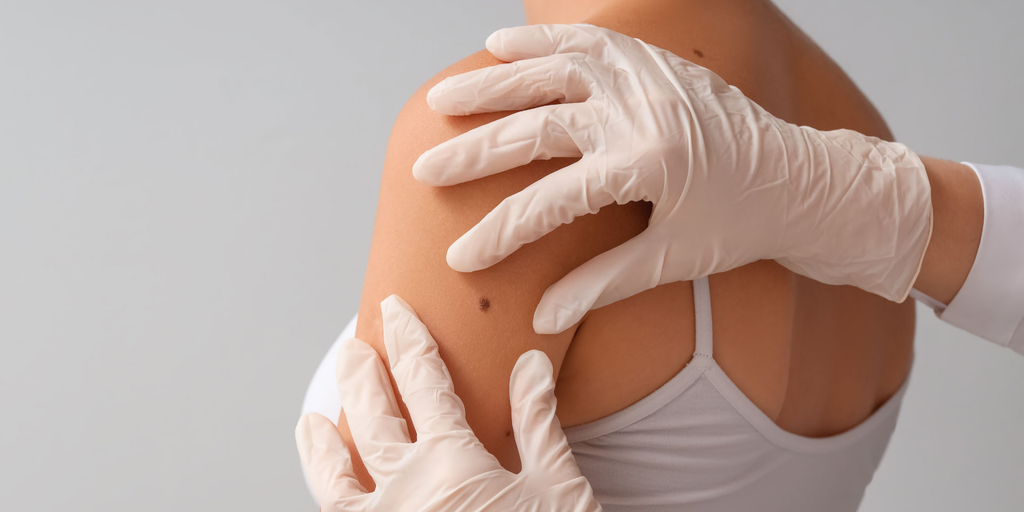
Finding a lump beneath or on the surface of the skin can raise questions about its cause, whether treatment is necessary, and what options are available. Similarly, an existing scar may cause discomfort, restrict movement, or affect a person’s confidence. Understanding when to seek medical advice is an important first step. excision of benign lumps is one treatment option for certain non-cancerous growths, while scar revision may help improve the appearance or function of selected scars. Both procedures require an individual assessment to determine whether surgery is appropriate.
In London, patients can seek advice from qualified medical professionals who assess skin lumps and scars before recommending treatment. The decision to proceed depends on several factors, including the nature of the lump, the characteristics of the scar, medical history, and personal expectations. Understanding these considerations can help patients prepare for a consultation and make informed decisions about their care.
Understanding Benign Lumps and When They Need Treatment
Benign lumps are non-cancerous growths that can develop in or beneath the skin. They may occur in different parts of the body and vary in size, shape, texture, and depth. Although many are harmless, some may require medical assessment or removal.
Common Types of Benign Lumps
Several types of non-cancerous lumps may be assessed for surgical removal, including:
- Lipomas: Soft growths made up of fatty tissue, usually found beneath the skin.
- Skin cysts: Closed sacs that may contain fluid, keratin, or other material.
- Dermatofibromas: Firm, usually harmless skin growths that can develop in different areas.
- Other benign growths: Certain non-cancerous lesions may require assessment to establish their nature and appropriate management.
Not every lump is suitable for immediate removal. A clinician may examine the area and recommend further investigations if the diagnosis is uncertain.
Signs That a Lump Should Be Assessed
A lump should be checked by a healthcare professional if it is new, growing, painful, changing in appearance, or associated with other unusual symptoms. A lump that becomes red, warm, swollen, or tender may also require medical attention, particularly if infection is suspected.
Rapid growth, changes in texture, or a lump that feels fixed to deeper tissues should not be ignored. Although these features do not automatically indicate a serious condition, they make proper assessment important.
When Is Benign Lump Excision Worth Considering?
Surgical removal may be recommended when a benign lump causes physical discomfort, interferes with everyday activities, becomes repeatedly inflamed, or creates cosmetic concerns. The decision should be based on a clinical assessment rather than appearance alone.
Physical Discomfort and Daily Activities
Some lumps can become uncomfortable when they rub against clothing, are exposed to pressure, or sit in areas that move frequently. For example, a growth near a waistband, shoulder strap, or joint may cause irritation during normal activities.
If a lump affects movement or causes repeated discomfort, a clinician may discuss whether removal is appropriate. The benefits should be weighed against potential risks, including bleeding, infection, recurrence, and scarring.
Cosmetic Concerns
Some people seek treatment because a visible lump affects their appearance or causes self-consciousness. This may be particularly relevant when a growth appears on the face, neck, or another exposed area.
A consultation can help establish what removal may involve and whether the expected scar is likely to be acceptable. Patients should understand that removing a lump does not always result in completely unnoticeable skin.
Changes in Size or Appearance
A lump that changes over time should be assessed before any cosmetic procedure is planned. A clinician may recommend imaging, a biopsy, or other investigations depending on its location and characteristics.
If a lump is removed, the tissue may be sent for histological examination when clinically indicated. This allows a laboratory to assess the tissue and help confirm the diagnosis.
Understanding Scar Revision and Its Potential Benefits
Scars form as part of the body’s natural healing process following injuries, operations, burns, or other skin damage. Their appearance varies according to the original wound, skin characteristics, healing response, and location.
Some scars become raised, wide, uneven, discoloured, or uncomfortable. Scar revision is a group of treatments intended to improve selected features of an existing scar.
Different Types of Scars
Understanding the type of scar can help determine which treatment options may be suitable.
- Hypertrophic scars: Raised scars that remain within the boundaries of the original wound.
- Keloid scars: Raised scars that extend beyond the original wound and may continue growing.
- Atrophic scars: Depressed scars that create an indentation in the skin.
- Contracture scars: Tight scars that may restrict movement, particularly after burns or significant injuries.
- Wide or irregular scars: Scars that have healed with an uneven shape or expanded appearance.
Each type may require a different approach. Not all scars are suitable for surgical revision, and some may respond better to non-surgical treatments.
When Should You Consider Scar Revision in London?
Patients may explore scar revision in London when an existing scar causes functional difficulties, persistent discomfort, or concerns about its appearance. A specialist assessment can help determine whether surgery or another treatment approach is more suitable.
Scars That Affect Movement or Comfort
Some scars become tight and restrict the movement of nearby skin or joints. This can happen when scar tissue forms across areas such as the neck, hands, elbows, or knees.
If a scar causes pulling, discomfort, or restricted movement, a medical assessment can establish whether treatment may improve function. The recommended approach depends on the scar’s location, depth, and underlying tissue involvement.
Scars That Remain Raised or Uneven
Raised, thickened, or irregular scars may be suitable for selected treatments. Depending on the scar type, options may include silicone products, injections, laser treatment, or surgical revision.
Surgery may involve removing or repositioning existing scar tissue and closing the wound using techniques intended to improve its shape or direction. However, a new scar will still form, and the final result cannot be guaranteed.
Scars Following Previous Surgery or Injury
Scars from earlier operations or injuries may heal with uneven edges, widening, or changes in skin texture. Some patients seek a consultation when a scar remains noticeable after the initial healing period.
A specialist can review the original wound, previous treatments, and current scar characteristics before discussing possible improvements.
What Happens During a Consultation?
A consultation is an opportunity to understand the condition, discuss treatment options, and establish realistic expectations. Patients should provide information about their medical history, previous procedures, allergies, medications, and any changes they have noticed.
Examination and Diagnosis
For a skin lump, the clinician may assess its size, location, mobility, consistency, and appearance. Further tests may be recommended if the diagnosis is uncertain.
For a scar, the assessment may include its thickness, colour, texture, flexibility, and effect on nearby movement. The clinician will also consider the patient’s tendency to develop raised or abnormal scars.
Discussing Treatment Options
Treatment recommendations should reflect the patient’s individual circumstances. Some benign lumps can be monitored rather than removed, while certain scars may be managed with non-surgical methods.
If surgery is recommended, the clinician should explain the procedure, anaesthesia, recovery expectations, possible complications, and likely scar formation. Patients should have the opportunity to ask questions before deciding.
Recovery and Aftercare Following Treatment
Recovery depends on the procedure, the size and location of the treated area, and individual healing characteristics. Minor lump removal may involve a relatively short recovery period, while more extensive procedures may require additional care.
Caring for the Treated Area
Patients should follow the aftercare instructions provided by their clinical team. These may include keeping the wound clean and dry as advised, changing dressings when instructed, avoiding unnecessary pressure, and attending follow-up appointments.
Signs such as increasing redness, worsening pain, discharge, fever, or unusual swelling should be reported promptly to a healthcare professional.
Allowing Scars Time to Mature
Scars continue changing for months after an operation. Their colour, thickness, and texture may gradually improve as the healing process progresses.
Patients should avoid judging the final appearance too early. Following wound-care guidance and protecting healing skin from excessive sun exposure can support recovery.
Making an Informed Decision About Treatment
Deciding whether to undergo benign lump excision or scar revision involves more than considering appearance. Patients should understand the diagnosis, the reason for treatment, alternative options, potential complications, and expected recovery.
Benign lump removal may be appropriate when a growth causes discomfort, repeated inflammation, functional difficulties, or cosmetic concerns. Scar revision may be worth discussing when an existing scar affects movement, causes discomfort, or remains a concern after healing.
Ultimately, the most appropriate treatment depends on an individual clinical assessment. Consulting a qualified specialist in London can help patients understand their options and decide whether surgery is suitable for their needs.
Conclusion
Benign lumps and noticeable scars can affect people in different ways, from physical irritation to concerns about appearance and movement. While some conditions can be monitored or managed without surgery, others may benefit from a specialist assessment and an appropriate treatment plan.
Understanding the reasons for removal, the different scar treatment options, and the possible outcomes can help patients approach consultations with greater confidence. A careful discussion with a qualified medical professional is the best way to establish whether benign lump excision or scar revision is appropriate and what patients can realistically expect from treatment.
How to Decide If You Need Penile Enlargement or a P-Shot in Bournemouth Based on Your Goals
How to Recover Smoothly From a Deep Plane Neck Lift and Midface Lift Surgery Together
How Head and Neck Surgery Specialists Use Sialendoscopy for Diagnosis

Meet Lisa Ann Mudd, Sydney Sweeney’s Mother and Former Attorney

Jason Lawson: The Story of Hayley Mills’ Son

Nina Weiss: All You Need to Know About Manuel Neuer’s Ex-Wife
-
 News3 months ago
Meet Lisa Ann Mudd, Sydney Sweeney’s Mother and Former Attorney
-
 Celebrity4 months ago
Jason Lawson: The Story of Hayley Mills’ Son
-
 Celebrity4 months ago
Nina Weiss: All You Need to Know About Manuel Neuer’s Ex-Wife
-

 Celebrity4 months ago
Sandra Davenport: The Untold Story of Jim Belushi’s First Marriage
-

 News3 months ago
Who Is Rebecca Dyer? Meet David Foster’s Ex-Wife
-

 Biographies3 months ago
Who is Thomas Earle? All About Alix Earle’s Father
-

 Celebrity4 months ago
Jack Scarborough: Everything About Joe Scarborough’s Son
-

 Celebrity4 months ago
Debra Rodman: Inside the Life of Dennis Rodman’s Powerful Sister