
Health
What to Understand About Scarring When Choosing Closed Rhinoplasty as Your Rhinoplasty in London Procedure


When considering cosmetic surgery to refine facial harmony, understanding how incision placement affects scarring remains a top priority. Patients researching closed rhinoplasty in London often seek a refined contour without visible marks across the columella. While every surgical procedure leaves some form of scar tissue, internal approaches conceal these marks within the nostrils. Choosing rhinoplasty in London requires a clear understanding of how different techniques handle tissue modifications, skin elasticity, and structural support. This guide breaks down the reality of post-operative scarring, helping prospective patients evaluate whether an endonasal approach suits their individual anatomical goals and aesthetic expectations.
Understanding Surgical Incisions in Nasal Reshaping
Surgical alterations to the nasal framework necessitate incisions to access the underlying cartilage and bone. In traditional open procedures, a small external cut is made across the columella—the tissue bridge separating the nostrils. In contrast, the endonasal or closed approach places all cuts entirely inside the nasal cavities.
Internal vs External Micro-Trauma
Because structural modifications occur internally, patients undergoing closed rhinoplasty in London avoid external scarring altogether. Inside the mucosal lining, small incisions allow the surgeon to adjust the dorsal hump, narrow the bridge, or reshape cartilage. These internal mucosal cuts heal remarkably well due to the rich blood supply within the nasal cavity.
How Mucosal Tissue Heals
Mucosal lining possesses distinct biological properties compared to external skin. It regenerates rapidly, reducing the likelihood of thick keloid or hypertrophic scar formation. While internal scar tissue does form during structural healing, it remains hidden from external view. Managing this internal healing process involves following strict post-operative cleansing routines and keeping the nasal passages moist with saline sprays. By understanding how mucosal tissue repairs itself, patients better appreciate why internal techniques offer a subtle, discreet recovery process.
Why Scarring Differs Between Open and Closed Procedures
The primary distinction between surgical approaches lies in the visibility and location of scar tissue. An open procedure provides direct visual access to the nasal structure by lifting the skin envelope, which requires an external columellar incision. Although skilled surgeons close this incision with fine sutures, a subtle external line remains.
Minimising External Marks
For individuals considering rhinoplasty in London, the closed approach eliminates external cut marks entirely. The absence of a columellar scar proves particularly advantageous for patients prone to hyperpigmentation or thick scarring. Because no skin is severed across the central partition, the external blood flow and lymphatic drainage remain largely undisturbed.
Impact on Post-Operative Swelling
Preserving the natural lymphatic pathways leads to another crucial benefit: reduced protracted swelling. External incisions interrupt lymphatic channels, often prolonging tip oedema for twelve to eighteen months. Internal incisions limit this disruption, allowing tissues to settle into their final position more rapidly. Consequently, the internal healing trajectory is smoother, letting patients appreciate their revised contours without waiting out extended external recovery phases.
Factors Influencing Scar Maturation and Healing Quality
Even when incisions remain internal, individual biological factors heavily influence how scar tissue develops and matures. Genetics, skin thickness, age, and lifestyle habits all play pivotal roles in tissue repair.
The Role of Skin Elasticity and Thickness
Patients with thicker nasal skin naturally produce more subcutaneous collagen during recovery. While this can cushion internal modifications, excessive scar tissue development beneath the skin might obscure fine tip definition. Conversely, thin skin reveals underlying structural changes immediately but provides less coverage for minor irregularities.
Lifestyle and Nutritional Factors
Post-surgical recovery demands proper physical support. Smoking severely constricts blood vessels, restricting oxygen delivery to healing tissue and increasing the risk of poor scar formation. Maintaining optimal hydration and consuming balanced nutrients—especially vitamin C and zinc—supports healthy cellular turnover. Choosing a qualified practitioner for rhinoplasty in London ensures patients receive detailed pre- and post-operative instructions tailored to their specific skin type, helping to prevent excess internal fibrous tissue formation.
Managing Internal Scar Tissue During Recovery
Proper post-operative management ensures internal scar tissue settles smoothly without affecting breathing or changing the aesthetic outline. During the first few weeks after surgery, the body initiates an inflammatory response, building collagen fibers to reinforce the modified cartilage and bone structure.
Adhering to Post-Operative Guidelines
Surgeons typically prescribe gentle saline irrigations and antibiotic ointments to keep internal incision lines soft and clean. Preventing crusting inside the nostrils allows the mucosal lining to re-epithelialise smoothly, reducing the risk of adhesions—where scar tissue bridges across internal nasal walls.
Steroid Injections and Tissue Softening
In cases where localised internal swelling or dense scar tissue persists around the nasal tip, targeted corticosteroid injections help. These minor treatments soften firm collagen bundles and encourage uniform contraction. Regular follow-up consultations allow the surgical team to monitor internal healing closely, making timely interventions if scar tissue begins to pull on the flexible lower lateral cartilages.
Selecting the Right Surgical Approach for Your Anatomy
While avoiding external scarring makes endonasal surgery highly appealing, it is not suitable for every cosmetic or functional goal. The decision between open and closed techniques depends on the complexity of structural changes required.
Assessing Anatomical Suitability
Minor to moderate dorsal hump reduction, subtle bridge narrowing, and slight tip adjustments respond exceptionally well to internal access. However, complex revision surgeries or severe tip asymmetry often require the maximum visibility provided by an open incision.
Consulting a Specialist
During an initial consultation for closed rhinoplasty in London, a surgeon evaluates nasal architecture, airway patency, and skin envelope. Discussing scar expectations openly ensures realistic goals. A skilled specialist balances scar minimisation with precise structural control to achieve natural, long-lasting results.
Final Thoughts
Choosing an endonasal approach for nasal reshaping offers significant advantages, particularly regarding post-operative scar management. By concealing all incision lines inside the nostrils, this method preserves external skin integrity while reducing recovery time and swelling. However, achieving superior aesthetic outcomes requires balancing scar avoidance with individual anatomical needs. Working alongside an experienced surgical team ensures tailored care from initial evaluation through complete tissue maturation. Understanding how internal healing works empowers patients to make confident decisions about their cosmetic journey, securing a refined, natural result without visible external marks.
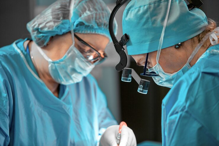
Salivary gland problems can cause discomfort, swelling, and recurring pain, particularly when saliva cannot flow normally through the ducts. For patients experiencing these symptoms, consulting London head and neck surgery specialists can help identify the underlying cause and determine an appropriate treatment approach. One diagnostic technique used in selected cases is sialendoscopy, a minimally invasive procedure that allows specialists to examine the inside of the salivary gland ducts.
Sialendoscopy provides a direct view of the duct system, helping clinicians investigate conditions such as salivary stones, duct narrowing, and inflammation. Unlike conventional imaging alone, this technique allows specialists to inspect the affected area using a small endoscope.
Understanding how the procedure works, when it may be recommended, and what patients can expect can help individuals prepare for a consultation with a qualified specialist.
What Is Sialendoscopy?
Sialendoscopy is a minimally invasive technique used to examine the ducts that carry saliva from the salivary glands into the mouth. It involves inserting a very thin endoscope equipped with a light and camera into a natural opening of the salivary duct.
The instrument allows the specialist to view the duct’s internal structure and identify abnormalities that may be responsible for a patient’s symptoms.
Understanding the Salivary Glands
The body has three major pairs of salivary glands:
- Parotid glands: Located in front of and below the ears.
- Submandibular glands: Found beneath the lower jaw.
- Sublingual glands: Situated beneath the tongue.
These glands produce saliva, which helps lubricate the mouth, support swallowing, and begin the digestive process. Saliva travels through small ducts before entering the oral cavity.
When a duct becomes blocked or narrowed, saliva may accumulate inside the gland. This can lead to swelling, discomfort, and sometimes infection.
How the Endoscope Works
The endoscope used during sialendoscopy is designed to pass through the natural salivary duct opening. Its small camera transmits images to a monitor, allowing the specialist to examine the duct’s interior.
Depending on the clinical findings, the procedure may also allow the specialist to investigate an obstruction more closely or perform certain treatments during the same session.
Why Specialists Use Sialendoscopy for Diagnosis
Salivary gland symptoms can have several possible causes. Swelling around the jaw, discomfort during meals, and repeated episodes of inflammation may indicate an issue involving the salivary ducts.
Sialendoscopy helps specialists investigate these symptoms by providing a direct view of the duct system.
Identifying Salivary Stones
Salivary stones, also known as salivary calculi, are hardened deposits that develop within the salivary glands or their ducts. They can obstruct saliva flow and cause swelling or pain, particularly when eating.
During sialendoscopy, a specialist may be able to identify the location and appearance of a stone. This information can help determine whether it may be suitable for removal using an endoscopic technique or whether another approach is necessary.
Detecting Duct Narrowing
Salivary ducts can become narrowed due to inflammation, scarring, or other structural changes. This condition, known as duct stenosis, may restrict saliva movement and contribute to recurring symptoms.
Sialendoscopy allows the specialist to examine the affected section and assess the extent of the narrowing. The findings can help guide decisions about further investigation or treatment.
Investigating Recurrent Inflammation
Repeated salivary gland inflammation may be associated with blockages, duct abnormalities, or other underlying conditions.
Direct examination of the duct can provide additional information when symptoms continue despite initial management. However, sialendoscopy is not suitable for every patient, and the specialist must assess whether it is appropriate.
When Is Sialendoscopy Recommended?
Sialendoscopy may be recommended when a patient’s symptoms suggest an obstruction or abnormality within a salivary duct. It is particularly useful when further investigation is needed after a clinical examination or imaging assessment.
Patients experiencing recurring swelling, discomfort during meals, or repeated episodes of salivary gland inflammation may benefit from specialist evaluation.
Symptoms That May Require Investigation
Common symptoms associated with salivary duct problems include:
- Swelling around the cheeks, jaw, or beneath the tongue.
- Pain that becomes more noticeable during meals.
- Recurrent salivary gland infections.
- A feeling of pressure around the affected gland.
- Reduced saliva flow or persistent dry mouth.
These symptoms do not automatically indicate a condition that requires sialendoscopy. Other medical problems can produce similar signs, so an appropriate assessment is important.
The Role of Medical Imaging
Before recommending an endoscopic examination, specialists may request imaging tests such as ultrasound, computed tomography (CT), or magnetic resonance imaging (MRI).
These investigations can help identify stones, gland abnormalities, and other structural changes. The choice of imaging depends on the patient’s symptoms and clinical history.
Sialendoscopy may provide additional information when imaging does not fully explain the problem or when direct examination of the duct is required.
How Sialendoscopy in London Supports Specialist Assessment
Access to specialist evaluation is important when salivary gland symptoms are persistent or recurring. Patients researching sialendoscopy in London can learn more about the procedure and discuss whether it may be suitable for their particular condition.
A specialist assessment usually begins with a discussion of symptoms, medical history, and previous investigations. The clinician may examine the affected area and review any available imaging before recommending further tests or treatment.
Preparing for the Procedure
Preparation depends on the planned procedure and the type of anaesthesia required. Patients may receive instructions about eating, drinking, medication, and arrangements for travelling home.
It is important to disclose existing medical conditions, allergies, and prescribed medication during the consultation. Patients should also ask the specialist about expected recovery time and any restrictions following the procedure.
What Happens During Sialendoscopy?
The procedure may be performed under local or general anaesthesia, depending on the clinical circumstances.
Once the patient is appropriately prepared, the specialist gently introduces the endoscope through the salivary duct opening. The camera provides images of the duct, allowing the clinician to inspect its internal lining and identify abnormalities.
If a stone or narrowing is found, the specialist may discuss whether treatment can be performed during the same procedure. The exact approach depends on the location, size, and nature of the problem.
Benefits and Limitations of Sialendoscopy
Sialendoscopy offers several potential advantages for selected patients, although it is not appropriate for every salivary gland condition.
Direct Visual Examination
One of its main advantages is the ability to examine the inside of the salivary duct directly. This can help specialists identify abnormalities that may not be fully characterised through other investigations.
Minimally Invasive Approach
Because the endoscope is introduced through the natural duct opening, the procedure may avoid the need for larger external incisions in suitable cases.
It can also allow certain obstructions to be treated while preserving the affected salivary gland. However, treatment outcomes depend on the individual condition and the findings during the procedure.
Possible Risks and Complications
Although generally considered minimally invasive, sialendoscopy carries potential risks. These may include temporary swelling, discomfort, bleeding, infection, or injury to the duct.
In some cases, the specialist may be unable to complete the examination or remove an obstruction. Further treatment may then be required.
Patients should discuss the possible benefits, limitations, and risks with their specialist before proceeding.
Recovery and Follow-Up After Sialendoscopy
Recovery varies according to the procedure performed, the patient’s general health, and whether any additional treatment was necessary.
Some patients experience mild discomfort or swelling for a short period. The specialist may provide instructions about oral hygiene, hydration, medication, and dietary adjustments.
Follow-up appointments allow the clinical team to review recovery and assess whether symptoms have improved. Patients should seek medical advice if they experience increasing pain, significant swelling, fever, or other concerning symptoms.
Conclusion
Sialendoscopy is a useful diagnostic technique for investigating selected salivary gland and duct conditions. By providing a direct view of the duct system, it can help specialists identify salivary stones, narrowing, and other abnormalities that may contribute to recurring symptoms.
For patients experiencing persistent salivary gland problems, a consultation with an experienced head and neck specialist can help establish whether sialendoscopy or another investigation is appropriate. Understanding the procedure, its potential benefits, and its limitations allows patients to make informed decisions about their care.
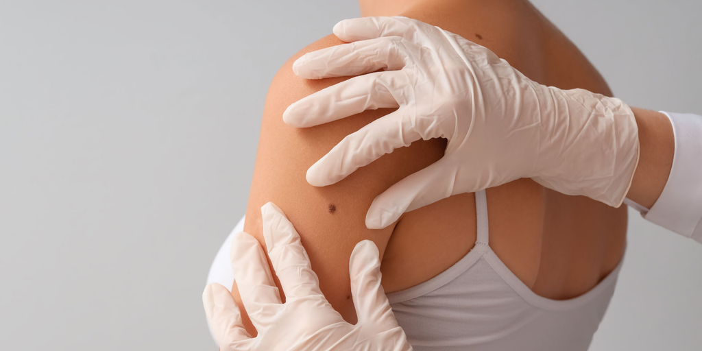
Finding a lump beneath or on the surface of the skin can raise questions about its cause, whether treatment is necessary, and what options are available. Similarly, an existing scar may cause discomfort, restrict movement, or affect a person’s confidence. Understanding when to seek medical advice is an important first step. excision of benign lumps is one treatment option for certain non-cancerous growths, while scar revision may help improve the appearance or function of selected scars. Both procedures require an individual assessment to determine whether surgery is appropriate.
In London, patients can seek advice from qualified medical professionals who assess skin lumps and scars before recommending treatment. The decision to proceed depends on several factors, including the nature of the lump, the characteristics of the scar, medical history, and personal expectations. Understanding these considerations can help patients prepare for a consultation and make informed decisions about their care.
Understanding Benign Lumps and When They Need Treatment
Benign lumps are non-cancerous growths that can develop in or beneath the skin. They may occur in different parts of the body and vary in size, shape, texture, and depth. Although many are harmless, some may require medical assessment or removal.
Common Types of Benign Lumps
Several types of non-cancerous lumps may be assessed for surgical removal, including:
- Lipomas: Soft growths made up of fatty tissue, usually found beneath the skin.
- Skin cysts: Closed sacs that may contain fluid, keratin, or other material.
- Dermatofibromas: Firm, usually harmless skin growths that can develop in different areas.
- Other benign growths: Certain non-cancerous lesions may require assessment to establish their nature and appropriate management.
Not every lump is suitable for immediate removal. A clinician may examine the area and recommend further investigations if the diagnosis is uncertain.
Signs That a Lump Should Be Assessed
A lump should be checked by a healthcare professional if it is new, growing, painful, changing in appearance, or associated with other unusual symptoms. A lump that becomes red, warm, swollen, or tender may also require medical attention, particularly if infection is suspected.
Rapid growth, changes in texture, or a lump that feels fixed to deeper tissues should not be ignored. Although these features do not automatically indicate a serious condition, they make proper assessment important.
When Is Benign Lump Excision Worth Considering?
Surgical removal may be recommended when a benign lump causes physical discomfort, interferes with everyday activities, becomes repeatedly inflamed, or creates cosmetic concerns. The decision should be based on a clinical assessment rather than appearance alone.
Physical Discomfort and Daily Activities
Some lumps can become uncomfortable when they rub against clothing, are exposed to pressure, or sit in areas that move frequently. For example, a growth near a waistband, shoulder strap, or joint may cause irritation during normal activities.
If a lump affects movement or causes repeated discomfort, a clinician may discuss whether removal is appropriate. The benefits should be weighed against potential risks, including bleeding, infection, recurrence, and scarring.
Cosmetic Concerns
Some people seek treatment because a visible lump affects their appearance or causes self-consciousness. This may be particularly relevant when a growth appears on the face, neck, or another exposed area.
A consultation can help establish what removal may involve and whether the expected scar is likely to be acceptable. Patients should understand that removing a lump does not always result in completely unnoticeable skin.
Changes in Size or Appearance
A lump that changes over time should be assessed before any cosmetic procedure is planned. A clinician may recommend imaging, a biopsy, or other investigations depending on its location and characteristics.
If a lump is removed, the tissue may be sent for histological examination when clinically indicated. This allows a laboratory to assess the tissue and help confirm the diagnosis.
Understanding Scar Revision and Its Potential Benefits
Scars form as part of the body’s natural healing process following injuries, operations, burns, or other skin damage. Their appearance varies according to the original wound, skin characteristics, healing response, and location.
Some scars become raised, wide, uneven, discoloured, or uncomfortable. Scar revision is a group of treatments intended to improve selected features of an existing scar.
Different Types of Scars
Understanding the type of scar can help determine which treatment options may be suitable.
- Hypertrophic scars: Raised scars that remain within the boundaries of the original wound.
- Keloid scars: Raised scars that extend beyond the original wound and may continue growing.
- Atrophic scars: Depressed scars that create an indentation in the skin.
- Contracture scars: Tight scars that may restrict movement, particularly after burns or significant injuries.
- Wide or irregular scars: Scars that have healed with an uneven shape or expanded appearance.
Each type may require a different approach. Not all scars are suitable for surgical revision, and some may respond better to non-surgical treatments.
When Should You Consider Scar Revision in London?
Patients may explore scar revision in London when an existing scar causes functional difficulties, persistent discomfort, or concerns about its appearance. A specialist assessment can help determine whether surgery or another treatment approach is more suitable.
Scars That Affect Movement or Comfort
Some scars become tight and restrict the movement of nearby skin or joints. This can happen when scar tissue forms across areas such as the neck, hands, elbows, or knees.
If a scar causes pulling, discomfort, or restricted movement, a medical assessment can establish whether treatment may improve function. The recommended approach depends on the scar’s location, depth, and underlying tissue involvement.
Scars That Remain Raised or Uneven
Raised, thickened, or irregular scars may be suitable for selected treatments. Depending on the scar type, options may include silicone products, injections, laser treatment, or surgical revision.
Surgery may involve removing or repositioning existing scar tissue and closing the wound using techniques intended to improve its shape or direction. However, a new scar will still form, and the final result cannot be guaranteed.
Scars Following Previous Surgery or Injury
Scars from earlier operations or injuries may heal with uneven edges, widening, or changes in skin texture. Some patients seek a consultation when a scar remains noticeable after the initial healing period.
A specialist can review the original wound, previous treatments, and current scar characteristics before discussing possible improvements.
What Happens During a Consultation?
A consultation is an opportunity to understand the condition, discuss treatment options, and establish realistic expectations. Patients should provide information about their medical history, previous procedures, allergies, medications, and any changes they have noticed.
Examination and Diagnosis
For a skin lump, the clinician may assess its size, location, mobility, consistency, and appearance. Further tests may be recommended if the diagnosis is uncertain.
For a scar, the assessment may include its thickness, colour, texture, flexibility, and effect on nearby movement. The clinician will also consider the patient’s tendency to develop raised or abnormal scars.
Discussing Treatment Options
Treatment recommendations should reflect the patient’s individual circumstances. Some benign lumps can be monitored rather than removed, while certain scars may be managed with non-surgical methods.
If surgery is recommended, the clinician should explain the procedure, anaesthesia, recovery expectations, possible complications, and likely scar formation. Patients should have the opportunity to ask questions before deciding.
Recovery and Aftercare Following Treatment
Recovery depends on the procedure, the size and location of the treated area, and individual healing characteristics. Minor lump removal may involve a relatively short recovery period, while more extensive procedures may require additional care.
Caring for the Treated Area
Patients should follow the aftercare instructions provided by their clinical team. These may include keeping the wound clean and dry as advised, changing dressings when instructed, avoiding unnecessary pressure, and attending follow-up appointments.
Signs such as increasing redness, worsening pain, discharge, fever, or unusual swelling should be reported promptly to a healthcare professional.
Allowing Scars Time to Mature
Scars continue changing for months after an operation. Their colour, thickness, and texture may gradually improve as the healing process progresses.
Patients should avoid judging the final appearance too early. Following wound-care guidance and protecting healing skin from excessive sun exposure can support recovery.
Making an Informed Decision About Treatment
Deciding whether to undergo benign lump excision or scar revision involves more than considering appearance. Patients should understand the diagnosis, the reason for treatment, alternative options, potential complications, and expected recovery.
Benign lump removal may be appropriate when a growth causes discomfort, repeated inflammation, functional difficulties, or cosmetic concerns. Scar revision may be worth discussing when an existing scar affects movement, causes discomfort, or remains a concern after healing.
Ultimately, the most appropriate treatment depends on an individual clinical assessment. Consulting a qualified specialist in London can help patients understand their options and decide whether surgery is suitable for their needs.
Conclusion
Benign lumps and noticeable scars can affect people in different ways, from physical irritation to concerns about appearance and movement. While some conditions can be monitored or managed without surgery, others may benefit from a specialist assessment and an appropriate treatment plan.
Understanding the reasons for removal, the different scar treatment options, and the possible outcomes can help patients approach consultations with greater confidence. A careful discussion with a qualified medical professional is the best way to establish whether benign lump excision or scar revision is appropriate and what patients can realistically expect from treatment.
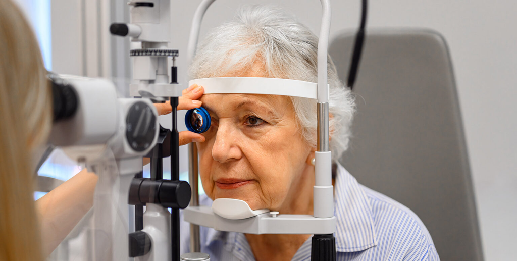
Cataract surgery and refractive lens exchange (RLE) are procedures that improve vision by replacing the eye’s natural lens with an artificial intraocular lens (IOL). Although they have different purposes, both require appropriate aftercare to support healing and reduce the risk of complications.
Following surgery, patients may experience temporary blurred vision, mild irritation, watery eyes or sensitivity to light. Taking appropriate precautions, using prescribed eye drops and following the surgeon’s instructions can help support recovery.
People undergoing cataract surgery in London or refractive lens exchange should understand which activities to avoid during recovery. This article explains ten important precautions and why they matter.
Understanding Recovery After Lens Replacement Surgery
Cataract surgery removes a cloudy natural lens, while refractive lens exchange removes the natural lens to correct refractive errors such as long-sightedness or short-sightedness.
Both procedures involve replacing the natural lens with an artificial alternative. Although patients may notice improvements in vision relatively quickly, the eye still needs time to heal.
Why Postoperative Care Matters
Postoperative care helps reduce the risk of infection, inflammation and other complications. Patients should follow their medication schedules, maintain good hygiene and attend scheduled appointments.
Recovery Can Vary Between Patients
Recovery depends on general eye health, the procedure performed and individual healing responses. Some patients resume daily activities within days, while others require additional time. Individual surgical instructions should always take priority over general advice.
1. Avoid Rubbing or Pressing Your Eyes
Patients should avoid rubbing or pressing the operated eye, even when it feels irritated, watery or uncomfortable. Touching the eye may interfere with healing and introduce bacteria.
Why Rubbing Can Be Harmful
Rubbing may increase infection risk and place unnecessary pressure on healing tissues. Patients should also avoid pressing their eyelids unless specifically instructed by their surgeon.
What to Do Instead
Use prescribed eye drops as directed and contact the clinic if irritation persists or worsens. Always wash your hands before administering medication and avoid touching the bottle tip against the eye.
2. Avoid Swimming and Hot Tubs During Early Recovery
Swimming pools, hot tubs, lakes and other bodies of water may expose the operated eye to microorganisms and irritants. Patients should avoid swimming until their surgeon confirms that it is safe.
Reduce Exposure to Contaminated Water
Pool chemicals may irritate the eye, while natural water sources can contain microorganisms. Hot tubs may also expose patients to bacteria.
Avoid swimming, diving and water-based recreational activities during the recommended restriction period.
Follow Your Surgeon’s Guidance
Ask when it is safe to resume swimming and using hot tubs. Showering is generally possible, but avoid allowing soap, shampoo or direct streams of water to enter the operated eye.
3. Avoid Strenuous Exercise and Heavy Lifting
Strenuous physical activity may need to be restricted during early recovery. Heavy lifting, intense workouts and sudden movements can place unnecessary strain on the body.
Activities to Limit Temporarily
Depending on medical advice, patients may need to avoid:
- Heavy weightlifting and resistance training.
- Running and vigorous aerobic exercise.
- High-intensity gym sessions.
- Contact sports and activities involving physical impact.
- Repeated bending or straining.
Return to Exercise Gradually
Light walking may be permitted relatively soon after surgery. However, patients should gradually resume demanding activities after receiving medical clearance. Stop exercising and seek advice if unusual discomfort or vision changes occur.
4. Avoid Getting Dust, Dirt and Chemicals in Your Eyes
Dust, smoke, debris and cleaning chemicals can irritate sensitive eyes during recovery. Patients should take additional care when completing household tasks or spending time outdoors.
Be Careful With Household Cleaning
Sweeping, vacuuming and dusting can release particles into the air. Strong cleaning products may also cause irritation. Where possible, ask someone else to complete these tasks during early recovery.
Protect Your Eyes Outdoors
Wind and airborne particles may cause discomfort. Suitable sunglasses can help reduce exposure to bright light and environmental irritants. Follow your surgeon’s recommendations regarding protective eyewear.
5. Avoid Driving Until You Are Cleared
Vision may remain blurry or fluctuate immediately after lens replacement surgery. Driving before eyesight has recovered sufficiently can put patients and other road users at risk.
Arrange Transport After Surgery
Arrange for someone to take you home after the procedure. Patients should not assume they can drive themselves, even when their vision appears relatively clear.
Check Your Vision Before Returning to the Road
Follow your surgeon’s advice and ensure that you meet the relevant legal eyesight requirements before driving. Glare, light sensitivity and difficulty judging distances may also affect driving ability.
6. Avoid Wearing Eye Makeup Too Soon
Mascara, eyeliner and eye shadow can introduce bacteria and small particles around the eyelids. Applying cosmetics too soon may increase irritation or infection risks.
Keep the Eye Area Clean
Avoid applying makeup close to the operated eye until your surgeon confirms that it is safe. Do not share cosmetics or use old and potentially contaminated products.
Ask When You Can Resume Your Routine
The appropriate time to restart makeup use varies. Follow the clinic’s instructions and use clean products and fresh applicators when you resume your routine.
7. Avoid Skipping Prescribed Eye Drops
Prescribed eye drops are an important part of postoperative care. Antibiotic and anti-inflammatory medications may help prevent infection and reduce inflammation.
Follow the Prescribed Schedule
Use medication exactly as instructed, even when your vision improves or discomfort disappears. Do not change the dosage, stop treatment early or introduce additional eye drops without professional advice.
Use Eye Drops Correctly
Wash and dry your hands before applying medication. Avoid touching the bottle tip against your eye, eyelids or other surfaces. Contact your clinic if you miss a dose or are unsure how to administer your medication.
8. Avoid Sleeping Without Following Eye Protection Advice
Some surgeons recommend wearing an eye shield while sleeping to prevent accidental rubbing or pressure during early recovery.
Why an Eye Shield May Be Recommended
People may unconsciously touch their eyes while asleep. An eye shield provides a physical barrier that helps reduce accidental contact.
Follow the Recommended Sleeping Position
Ask whether you should avoid sleeping on the operated side. Follow instructions about positioning, keeping the shield clean and how long it should be worn.
9. Avoid Ignoring Unusual Symptoms
Mild blurred vision, watery eyes and light sensitivity may occur during recovery. However, certain symptoms can indicate complications and require urgent assessment.
Recognise Warning Signs
Contact your eye care provider immediately if you experience:
- Sudden or significant vision loss.
- Severe or increasing eye pain.
- Worsening redness or discharge.
- New flashes of light or increased floaters.
- A curtain-like shadow across your vision.
- Increasing light sensitivity.
Sudden vision changes, severe pain or a curtain-like shadow require urgent medical attention.
Attend All Follow-Up Appointments
Follow-up appointments allow the surgeon to assess healing, check the artificial lens and identify potential complications. Attend every scheduled appointment, even when your vision appears to be improving.
10. Avoid Returning to Normal Activities Too Quickly
Patients may feel ready to resume their usual routines soon after surgery. However, returning to demanding activities too quickly may interfere with recovery.
Take Care With Work and Screen Time
Some patients can return to office work relatively soon, while others need additional time. Prolonged screen use may contribute to dry eyes or discomfort. Taking regular breaks and following recommended eye care instructions can help.
Give Yourself Time to Recover
Avoid comparing your recovery with someone else’s experience. Healing varies, so follow your surgeon’s recommendations before resuming work, exercise, travel and other activities.
Cataract Surgery and Refractive Lens Exchange: Are the Recovery Precautions Different?
Both procedures involve removing the natural lens and replacing it with an artificial intraocular lens. Consequently, many postoperative precautions are similar, although their treatment objectives differ.
Recovery After Cataract Surgery
Cataract surgery removes a cloudy natural lens that affects vision. Patients may notice improvements in visual clarity as the eye heals and adjusts to the artificial lens. The surgeon will monitor recovery and determine whether additional treatment or vision correction is necessary.
Recovery After Refractive Lens Exchange
Refractive lens exchange is generally performed to correct refractive errors rather than remove an existing cataract. Patients undergoing refractive lens exchange surgery in London should follow the same general principles of eye protection, medication use and postoperative monitoring.
Their surgeon can provide individual guidance about visual changes, recovery milestones and when normal activities can resume.
Conclusion
Recovery after cataract surgery and refractive lens exchange requires patience and careful attention to postoperative instructions. Avoiding eye rubbing, swimming, strenuous exercise, contaminated environments and premature returns to demanding activities can help protect the operated eye.
Patients should also use prescribed eye drops correctly, follow protective eye shield recommendations and attend all follow-up appointments. Driving and wearing eye makeup should only resume when the surgeon confirms that it is appropriate.
Sudden vision changes, severe pain, worsening redness and new flashes or floaters require urgent assessment. Following an individual recovery plan and communicating with an eye care professional can help patients manage healing safely and return to their usual activities at the appropriate time.
How Head and Neck Surgery Specialists Use Sialendoscopy for Diagnosis
How to Decide if You Need Benign Lump Excision and Scar Revision in London
What to Avoid After Cataract Surgery and Refractive Lens Exchange Surgery in London

Meet Lisa Ann Mudd, Sydney Sweeney’s Mother and Former Attorney

Jason Lawson: The Story of Hayley Mills’ Son

Nina Weiss: All You Need to Know About Manuel Neuer’s Ex-Wife
-
 News3 months ago
Meet Lisa Ann Mudd, Sydney Sweeney’s Mother and Former Attorney
-
 Celebrity4 months ago
Jason Lawson: The Story of Hayley Mills’ Son
-
 Celebrity4 months ago
Nina Weiss: All You Need to Know About Manuel Neuer’s Ex-Wife
-

 Celebrity4 months ago
Sandra Davenport: The Untold Story of Jim Belushi’s First Marriage
-

 News3 months ago
Who Is Rebecca Dyer? Meet David Foster’s Ex-Wife
-

 Biographies3 months ago
Who is Thomas Earle? All About Alix Earle’s Father
-

 Celebrity4 months ago
Jack Scarborough: Everything About Joe Scarborough’s Son
-

 Celebrity4 months ago
Debra Rodman: Inside the Life of Dennis Rodman’s Powerful Sister